
AXON
(Axonal BiomechaniX to Unveil Injury Mechanisms in Glaucomatous Optic Neuropathy)
Help us understand how the optic nerve responds to pressure and why some eyes may be more vulnerable to glaucoma.
Discover
Provide next-gen ophthalmic imaging platforms for biomarker discovery and oculomics research.
Translate
Serve as a translational sandbox for integrating and validating AI tools within real-world clinical workflows.
Educate
Advance patient understanding of AI in eye care through direct exposure and education.
Ultra-wide Field OCT

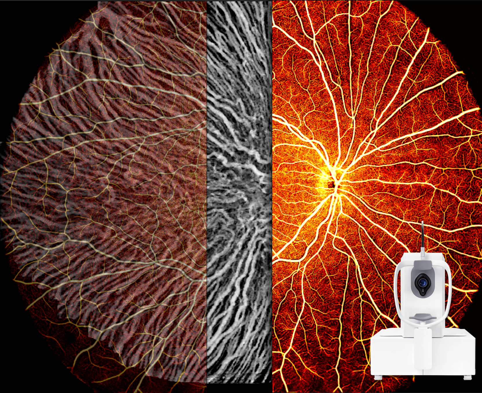
DREAM OCT provides ultra-wide-field, high-resolution 3D imaging of the retina, optic nerve head, choroid, and retinal blood vessels across an area of up to 26 × 21 mm in a single rapid scan.
Adaptive Optics OCT
m2OCT AO combines adaptive optics with depth-resolved OCT and angiography to reveal photoreceptors, retinal microvasculature, axon bundles, and other microscopic structures in three dimensions.
OCT Elastography
When combined with OCT, IOPstim safely and briefly increases intraocular pressure to reveal whether your ocular structures are mechanically robust or frail, helping identify biomechanical factors that may contribute to glaucoma risk.
Doppler Holography

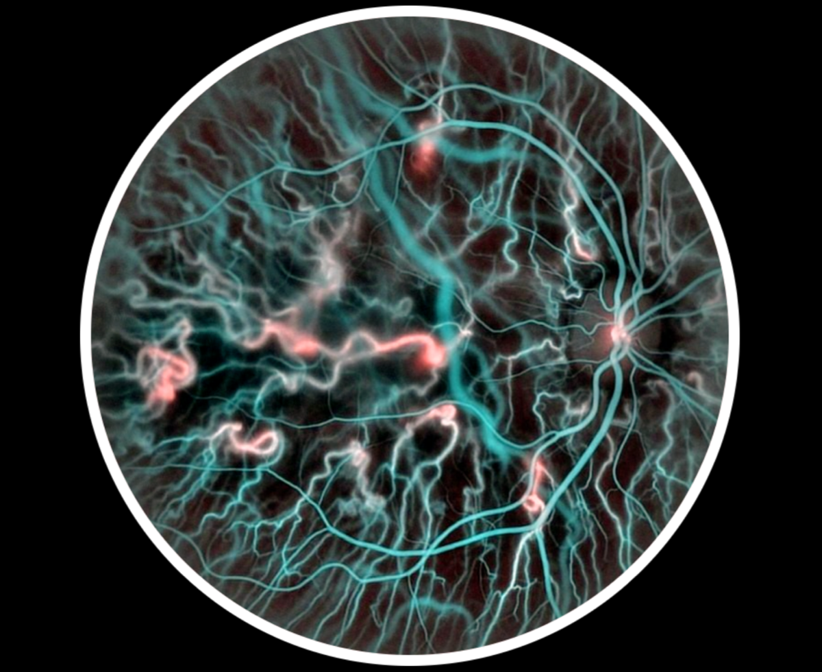
Doppler holography measures blood flow in the retina and optic nerve head, providing a functional assessment of how well oxygen and nutrients are delivered to these tissues.
Visible Light OCT
Aurora X4 uses visible-light OCT to provide ultra-high-resolution imaging of the retina and optic nerve head, while revealing tissue oxygenation and retinal microvascular function.
Spectral Domain OCT

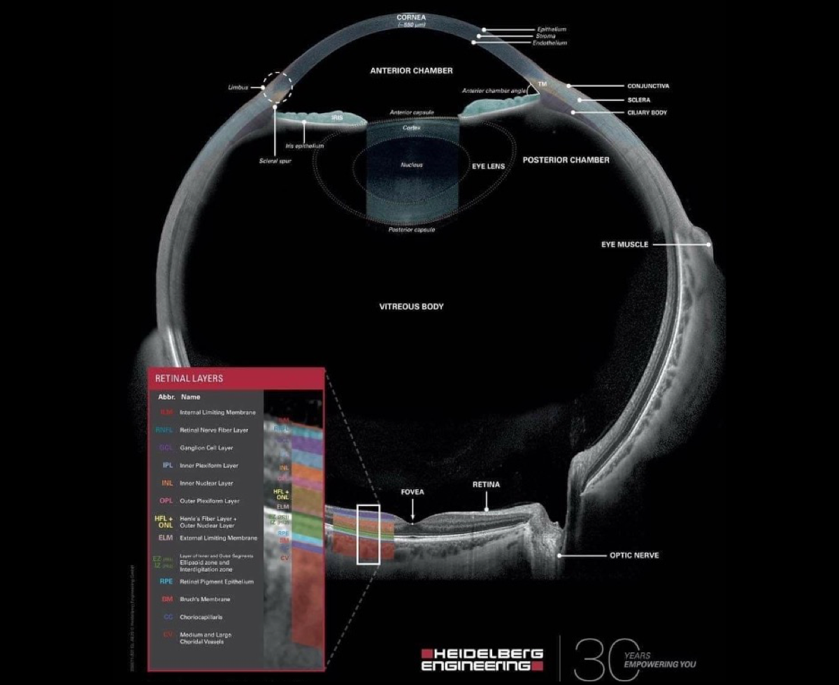
Spectralis is a standard clinical OCT system widely used in clinics worldwide, providing high-resolution imaging of the retina, optic nerve head, and choroid with precise eye tracking.
Adaptive Optics Camera

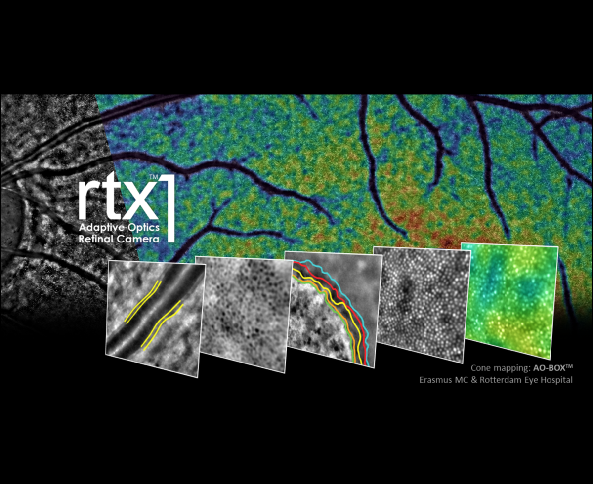
rtx1 provides en face, cellular-level imaging of the retina, enabling direct visualization and measurement of individual photoreceptors, retinal vessels, and microscopic retinal structures.
Glaucoma
Myopia
Optic neuritis
Ischemic optic neuropathy
Hereditary optic neuropathy
Diabetic retinopathy
Age-related macular degeneration
Retinal vascular disease
Optic disc drusen
Alzheimer's disease
Diabetes
Compressive optic neuropathy
Cardiovascular disease
Neurological disease
Clinical Imaging Studies
Help us build the next generation of eye care. By participating in one of our clinical imaging studies, you will see extraordinary images of your own eyes while helping us collect the high-quality data needed to develop robust and reliable AI.
(Axonal BiomechaniX to Unveil Injury Mechanisms in Glaucomatous Optic Neuropathy)
Help us understand how the optic nerve responds to pressure and why some eyes may be more vulnerable to glaucoma.
(Biobank at EmorY for Oculomics Using Next-Generation Imaging Devices)
Explore your eyes using our next-generation imaging technologies and help us discover new biomarkers of eye and systemic health.
Interested in participating?
Contact our team to learn moreInterested in visiting the Eagle Eye Suite, exploring our technologies, participating as a patient in research, or collaborating with our team? Get in touch with us to start the conversation.